Enhancing Cancer Diagnosis Through AI-Driven Technology
Written on
Chapter 1: Understanding Cancer Diagnosis
In the realm of cancer care, the biopsy is a crucial procedure often recommended by physicians after a physical examination or imaging suggests the possibility of cancer. This procedure involves extracting tissue from a suspicious area, which is then examined by a specialist known as a pathologist. Today, we will delve into the biopsy process and explore an innovative approach: utilizing artificial intelligence (AI) to enhance the speed and accuracy of cancer diagnoses.
The biopsy process is a key step in confirming a cancer diagnosis.
Section 1.1: The Basics of Biopsy
To establish a definitive cancer diagnosis, particularly for most types, a biopsy is generally required. The main types of biopsy include:
- Incisional Biopsy: Removal of a portion of the concerning tissue.
- Needle Biopsy: Extraction of a sample using a needle.
- Excisional Biopsy: Complete removal of the area of concern.
It's worth noting that cancer treatment is increasingly tailored to individual patients. The tissue obtained from a biopsy must yield sufficient material, not only for diagnosis but also for potential genetic testing.
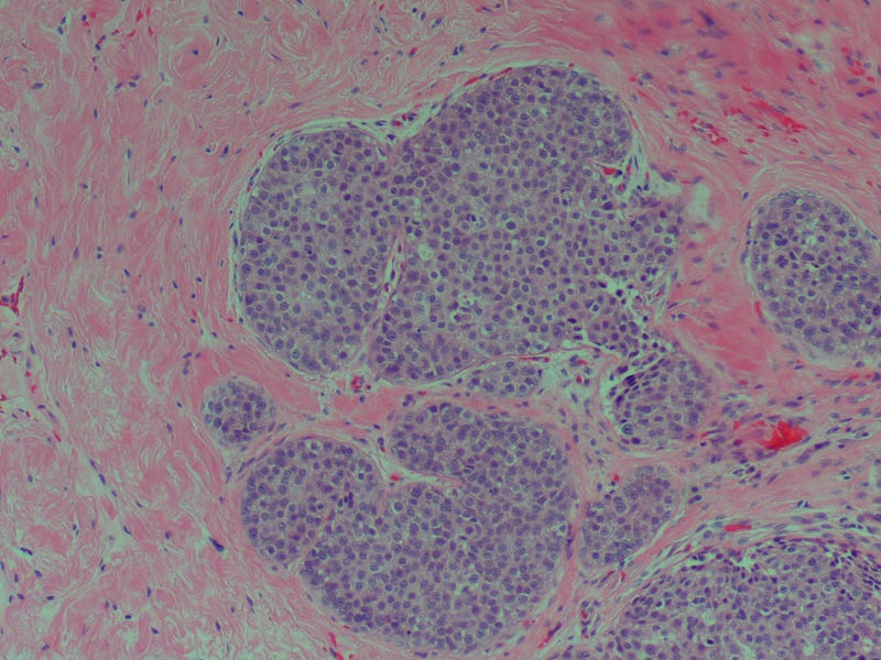
After the biopsy, the sample is sent to a pathologist who inspects the cells through a microscope. The pathologist then determines whether the tissue is benign (non-cancerous) or malignant (cancerous).
Subsection 1.1.1: Subjectivity in Diagnosis
Diagnosis can involve subjective interpretation. For instance, ductal carcinoma in situ (DCIS) is an early-stage form of breast cancer. When the cancer cells exhibit more aggressive characteristics (higher grade), diagnosing the condition is generally straightforward. However, lower-grade cases can pose a challenge. The spectrum of breast cancer ranges from normal cells to invasive cancer, with various intermediary stages.

Section 1.2: Errors in Pathology
Given the subtle differences between low-grade DCIS and atypical hyperplasia, diagnostic errors can occur. Such mistakes can have significant implications, as DCIS typically requires treatment while atypical ductal hyperplasia does not.
How frequently do pathologists misdiagnose breast cancer? Studies indicate they are proficient at identifying invasive breast cancer, but they face greater challenges with less severe conditions or normal tissue.
A comprehensive study involving over a hundred pathologists in the United States compared their diagnoses with those of three national experts. The findings revealed that:
- Pathologists accurately identified abnormal, pre-cancerous cells about 50% of the time, with follow-up typically involving monitoring and sometimes preventive medication.
- Approximately one-third of these cases were incorrectly diagnosed as non-problematic, while 17% were flagged as more suspicious or cancerous.
- In normal tissue samples, pathologists mistakenly identified 13% as suspicious.
Specifically for DCIS, 13% were misidentified as less severe, while 3% were misclassified as invasive cancer.

Chapter 2: The Future of Cancer Diagnosis
Recognizing that roughly 5% of cancer diagnoses are inaccurate, the U.S. Department of Defense has recently partnered with Google to enhance diagnostic precision. Google is leveraging neural networks and the Google Cloud Healthcare API to develop an AI system designed to assist pathologists.
The biopsy samples are anonymized, and once the AI is trained, Google aims to create a microscope that integrates augmented reality. This innovative device will provide pathologists with real-time data regarding the likelihood of malignancy in the examined cells.
If successful, this AI-driven approach will complement existing technologies used in cancer care. Pathologists may use AI to detect specific proteins, like HER2 in breast cancer, while radiologists can utilize AI for secondary evaluations of mammograms. The future of cancer diagnosis is rapidly advancing.
This first video discusses the groundbreaking use of machine learning in real-time cancer detection, highlighting how technology can revolutionize diagnostics.
The second video explores an AI-powered microscope that can assess cancer margins within minutes, showcasing the potential for rapid, accurate diagnostics.
I’m Dr. Michael Hunter. Thank you for joining me today.
Reference
Diagnostic concordance among pathologists interpreting breast biopsy specimens - PubMed
In this study of pathologists, in which diagnostic interpretation was based on a single breast biopsy slide, overall…
pubmed.ncbi.nlm.nih.gov